Method development
Tissue-based analyses
The analysis of the brain in neurological diseases or the affected tissue in cancer diseases has been the focus of our research for a long time, especially the elucidation of processes that have protective or disease-specific effects on cells. The isolation of specific cells from the respective tissue is a challenging task. Neurons in the central nervous system, for example, are highly branched, making it almost impossible to isolate neurons using traditional purification methods. One approach we use for our analyses is the LMD method, in which individual cells and cell components can be isolated from surrounding tissue. In this way, the proteome of degenerating neurons or cancer-affected tissue/cells can be specifically examined for changes using mass spectrometry.
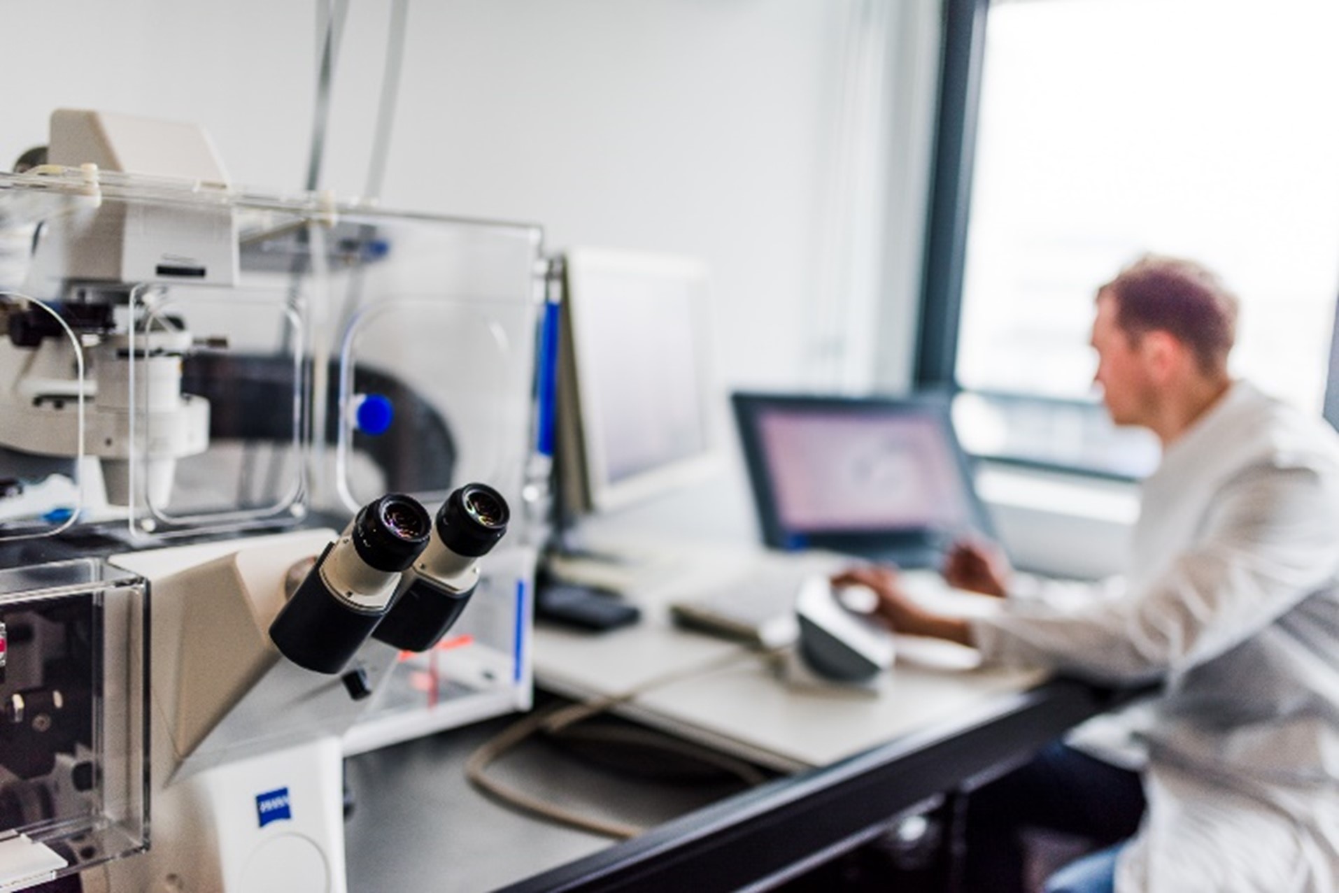
Figure 1: Analysis on the laser microdissection device.
For this purpose, the tissue is first cut ultra-thin, placed on slides, areas of interest are stained and then visualised with computer support and virtually marked. A laser beam travels around this virtual marking on the actual tissue section and catapults the tissue into a reaction vessel with another laser pulse. The basic prerequisite for the targeted enrichment of specific cells or tissue areas is visualisation. Here we use immunohistochemical methods with which we can stain specific proteins, but also the label-free spectroscopy-based tissue diagnostics developed by the Biospectroscopy competence area. In the field of neurodegenerative diseases, we have already succeeded in identifying disease-specific changes in the proteome of individual neuron populations and the pigment neuromelanin (REF). In the field of oncological diseases, we were able to perform proteomic analyses for the first time using FTIR/Raman imaging at spatial resolution on very precisely annotated but still untreated samples (Großerueschkamp et al Sci. Rep. 2017).
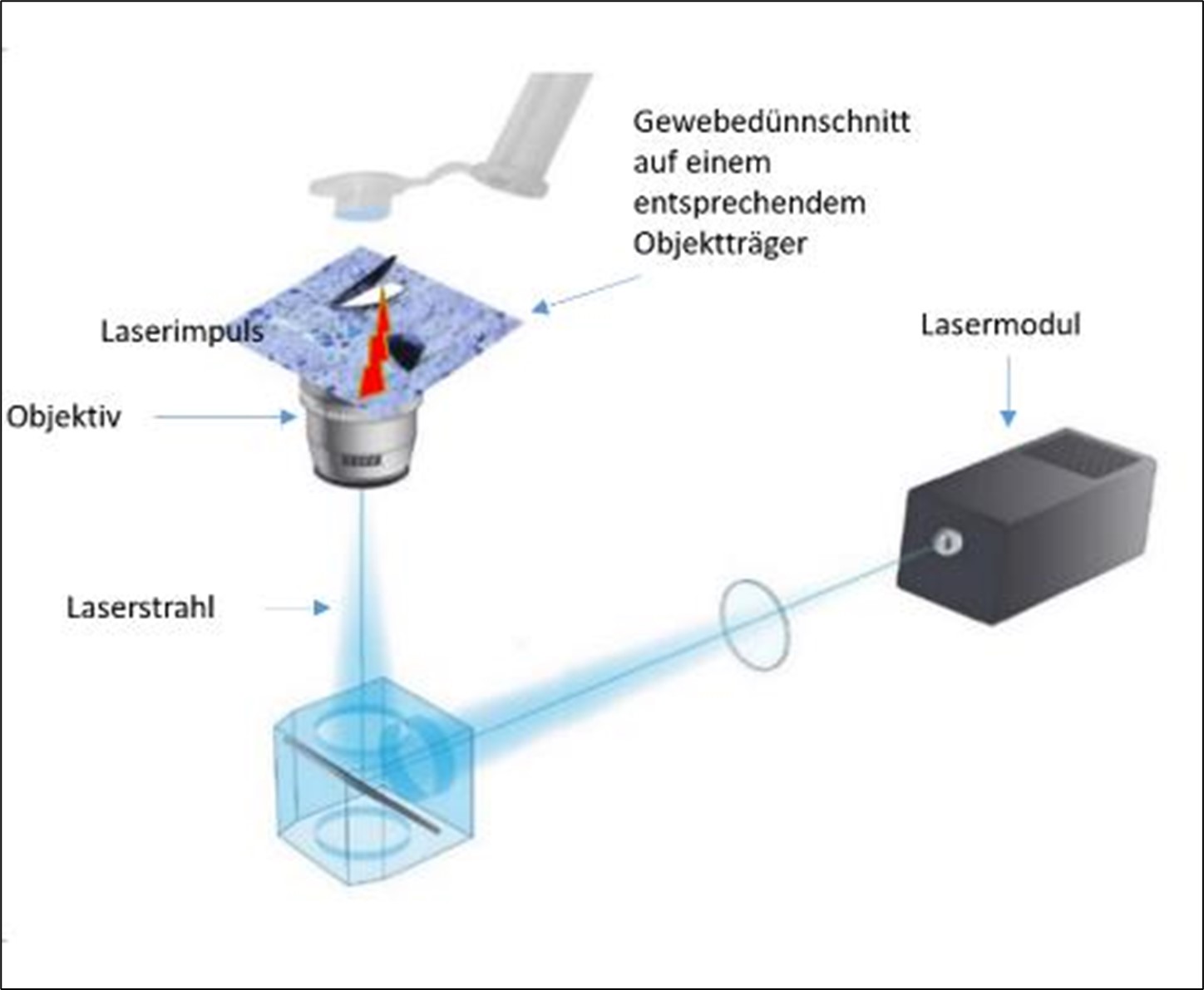
Figure 2: Schematic representation of laser microdissection.